


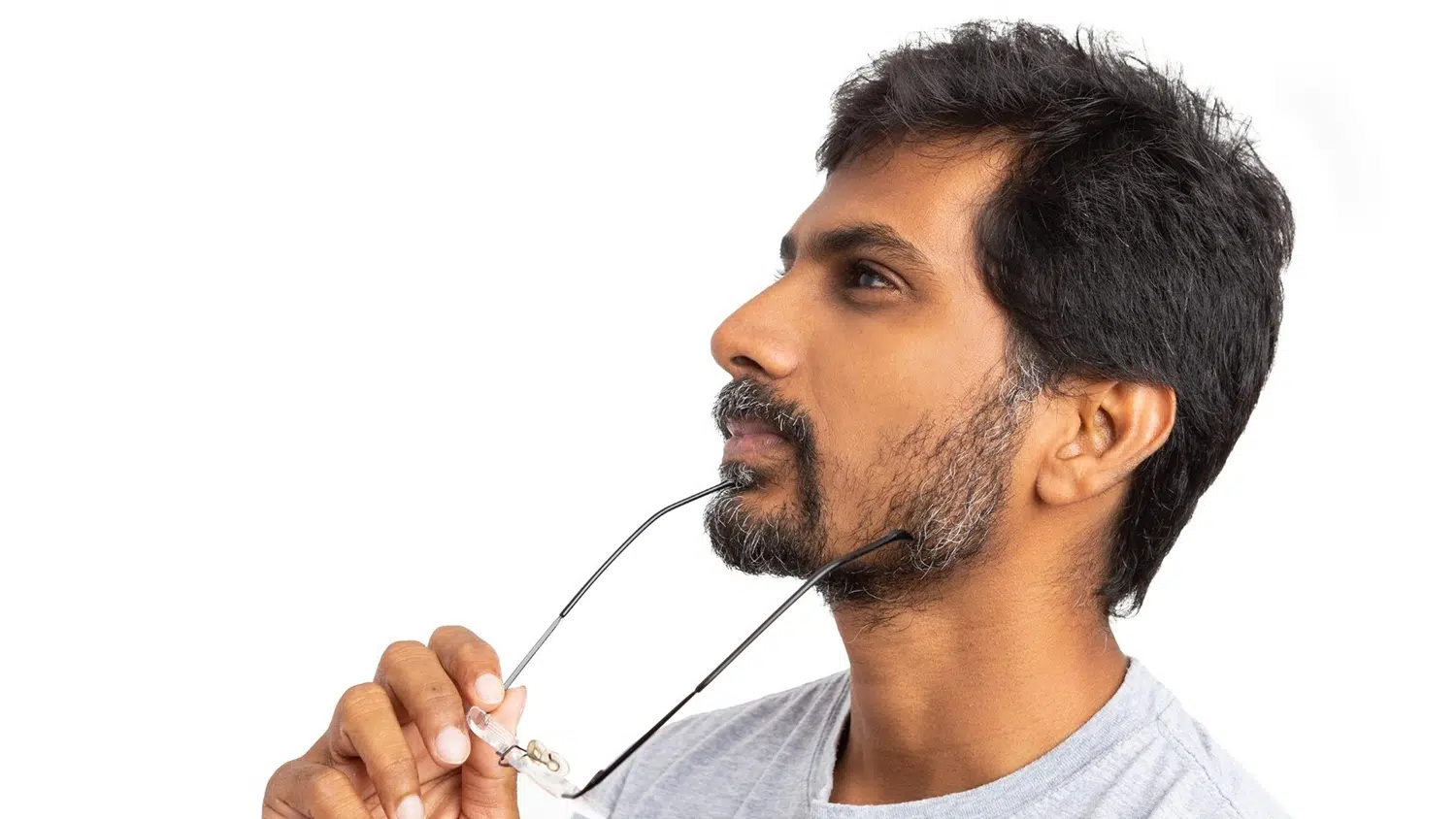
A person in Singapore whose glasses have not changed in ten years can still be developing changes at the back of the eye that only an examination will pick up. This is why highly short-sighted adults are advised to keep up regular eye checks for life, even when nothing feels wrong, and it applies just as much to those who have had laser surgery. That surgery corrects the blur, but it does not remove the underlying risk: it changes the focus of the eye, not its length. The most important thing to know is which warning signs mean act now. A sudden shower of floaters, flashes of light, or a dark curtain across your vision can signal a retinal tear, and these need same-day attention rather than a wait-and-see approach. This article explains what high myopia does to the eye over the years, which complications matter most, and the signs that separate a routine check from an emergency. Read on for the full picture.
By his forties, Mr Raj (not his real name) had made his peace with being very short-sighted. He had worn thick glasses since primary school, switched to contact lenses in his twenties, and long ago stopped thinking of it as anything more than an inconvenience. His prescription had held steady for years. His lenses worked. As far as he was concerned, his eyes had settled down and the story was over.
What he did not know was that a very short-sighted eye is never quite finished changing. Decades of myopia had left the back of his eyes stretched and thin, in a way no prescription could show and no contact lens could fix. The blur had stopped getting worse, so he assumed the risk had stopped too. The two are not the same thing.
He had also assumed that because he could see well with his lenses in, an eye check was something he could keep putting off. He was busy. Nothing hurt. Scrolling the news on the MRT each morning, he had a hundred more urgent things than sitting in a waiting room to be told what he already believed: that his eyes were simply short-sighted, as they had always been.
Then one evening, reading in bed, he noticed a scatter of tiny new floaters drifting across the vision in his left eye, and a flicker of light at the edge, like a camera flash that no one else could see. He blinked. It did not clear. Here is what those stretched, thinned eyes had been quietly setting up for years.
What Are the Main High Myopia Complications?

“The mistake I see most often is treating a stable prescription as a settled eye,” says Dr Allan Fong, an eye specialist in Singapore. “I describe it to patients as a balloon stretched thin. Once an eye has been blown up long and large by years of short-sightedness, the wall at the back stays fragile, even after the blurring stops changing. That eye needs watching for the rest of a person’s life.”
A large review of the evidence found that the risk of these sight-threatening problems climbs steeply as short-sightedness deepens, with high myopia carrying the greatest risk of all (Haarman et al., 2020).
Why Does a Stable Prescription Still Need Watching?
Laser refractive surgery can free a person from glasses by reshaping the front of the eye, but it does nothing to the length at the back, so the underlying risk travels with the patient regardless. This was the part that unsettled Mr Raj, who had assumed his contact lenses meant his eyes were as sturdy as anyone else’s.

What to watch for
- A short-sighted prescription that has been strong since childhood
- Being told at a past visit that your eyes are longer than average
- Assuming laser surgery has removed the need for future checks
Going years between eye examinations because nothing feels wrong
What Does High Myopia Do to the Back of the Eye?

Myopic macular degeneration matters because it affects the macula, the small central zone that lets you read, recognise faces across the table and see fine detail. Unlike the age-related form, it is driven by the mechanical stretching of a long eye rather than by age alone, and it is comparatively common in East Asian populations, including here in Singapore (Singapore Epidemiology of Eye Diseases Study).
Meanwhile, the stretched edges of the retina can develop thin patches and lattice-like weak points, which is often where tears begin. Mr Raj had none of the symptoms of central damage. His trouble was starting quietly, right at the edge.
What to watch for
- Straight lines that begin to look bent or wavy
- A blur or grey patch in the very centre of your sight
- Colours or fine detail that seem dimmer in one eye
- Changes that creep in gradually over weeks or months
What Are the Warning Signs of Retinal Detachment?
A retinal tear is the emergency at the sharp end of this condition, and it does not usually hurt, which is exactly why it is missed. The signs are visual, not painful: a shower of new floaters that will not clear, arcs or flashes of light at the edge of vision, or a shadow creeping in from one side like a curtain being drawn.
This is what Mr Raj noticed that night. If you are very short-sighted and any of these appear, treat it as urgent and seek help the same day rather than waiting to see if it passes. The Angel Eye Retinal Detachment page sets out these warning signs in more detail (National Eye Institute).
What to watch for
- A sudden burst of new floaters in one eye
- Repeated flashes of light, often at the outer edge
- A dark curtain or shadow moving across your vision
- A sudden drop in vision, with or without the above
Frequently Asked Questions About Cataracts
Is high myopia dangerous even if my prescription is stable?
Yes. A stable prescription only means the blur has stopped changing. The eye can still be long and the retina stretched and thin, which keeps the risk of high myopia complications such as retinal detachment and myopic macular degeneration in place for life. Regular checks matter even when vision feels fine.
What are the warning signs of retinal detachment?
The main warning signs are a sudden increase in floaters, flashes of light at the edge of vision, and a dark shadow or curtain moving across the eye. They are usually not painful. In a highly short-sighted person in Singapore, these signs call for same-day assessment to protect central vision.
How often should highly short-sighted adults have their eyes checked?
Most highly short-sighted adults benefit from a dilated eye examination at least once a year, or sooner if symptoms appear. Regular monitoring lets an eye doctor track the stretched retina over time and catch changes early, long before they threaten the centre of your sight.
When to Seek Help
A sudden shower of new floaters in one eye
Flashes of light, particularly at the edge of your vision
A shadow or curtain moving across your field of view
A sudden change in the clarity of your central vision
Straight lines that have started to look bent or distorted
At Angel Eye & Cataract Centre, Dr Allan Fong and the team provide thorough eye examinations for short-sighted adults in Singapore, including dilated checks of the peripheral and central retina and detailed imaging of the back of the eye. As an eye specialist, Dr Allan Fong assesses the structure of highly myopic eyes, monitors for change over time, and explains what each finding means for your long-term vision. Where warning signs suggest a retinal tear, the clinic can arrange prompt assessment.
If you have lived with strong short-sightedness for years, your eyes deserve a proper look even when nothing feels wrong. Booking a comprehensive eye examination with an eye specialist at Angel Eye & Cataract Centre is a simple way to stay ahead of high myopia complications and to protect the vision you rely on every single day.
Medical References
- Haarman AEG, Enthoven CA, Tideman JWL, Tedja MS, Verhoeven VJM, Klaver CCW. The Complications of Myopia: A Review and Meta-Analysis. Invest Ophthalmol Vis Sci. 2020;61(4):49. doi:10.1167/iovs.61.4.49. PMID: 32347918; PMCID: PMC7401976.
- Ohno-Matsui K, Wu PC, Yamashiro K, et al. IMI Pathologic Myopia. Invest Ophthalmol Vis Sci. 2021;62(5):5. doi:10.1167/iovs.62.5.5.
- Flitcroft DI. The complex interactions of retinal, optical and environmental factors in myopia aetiology. Prog Retin Eye Res. 2012;31(6):622-660. doi:10.1016/j.preteyeres.2012.06.004.
- Morgan IG, Ohno-Matsui K, Saw SM. Myopia. Lancet. 2012;379(9827):1739-1748. doi:10.1016/S0140-6736(12)60272-4.
- Wong TY, Ferreira A, Hughes R, Carter G, Mitchell P. Epidemiology and disease burden of pathologic myopia and myopic choroidal neovascularization: an evidence-based systematic review. Am J Ophthalmol. 2014;157(1):9-25. doi:10.1016/j.ajo.2013.08.010.
- Majithia S, Tham YC, Chee ML, et al. Cohort Profile: The Singapore Epidemiology of Eye Diseases study (SEED). Int J Epidemiol. 2021;50(1):41-52. doi:10.1093/ije/dyaa238.
- Ludwig CA, Vail D, Al-Moujahed A, et al. Epidemiology of rhegmatogenous retinal detachment in commercially insured myopes in the United States. Sci Rep. 2023;13(1):9430. doi:10.1038/s41598-023-35520-x.
- Ohno-Matsui K, Lai TYY, Lai CC, Cheung CMG. Updates of pathologic myopia. Prog Retin Eye Res. 2016;52:156-187. doi:10.1016/j.preteyeres.2015.12.001.
- National Eye Institute. Retinal Detachment. National Institutes of Health. nei.nih.gov.
- American Academy of Ophthalmology. Nearsightedness: Myopia and High Myopia. aao.org.
- Haarman AEG, et al. Axial length and the risk of myopic maculopathy and visual impairment. Invest Ophthalmol Vis Sci. 2020;61(4):49.
